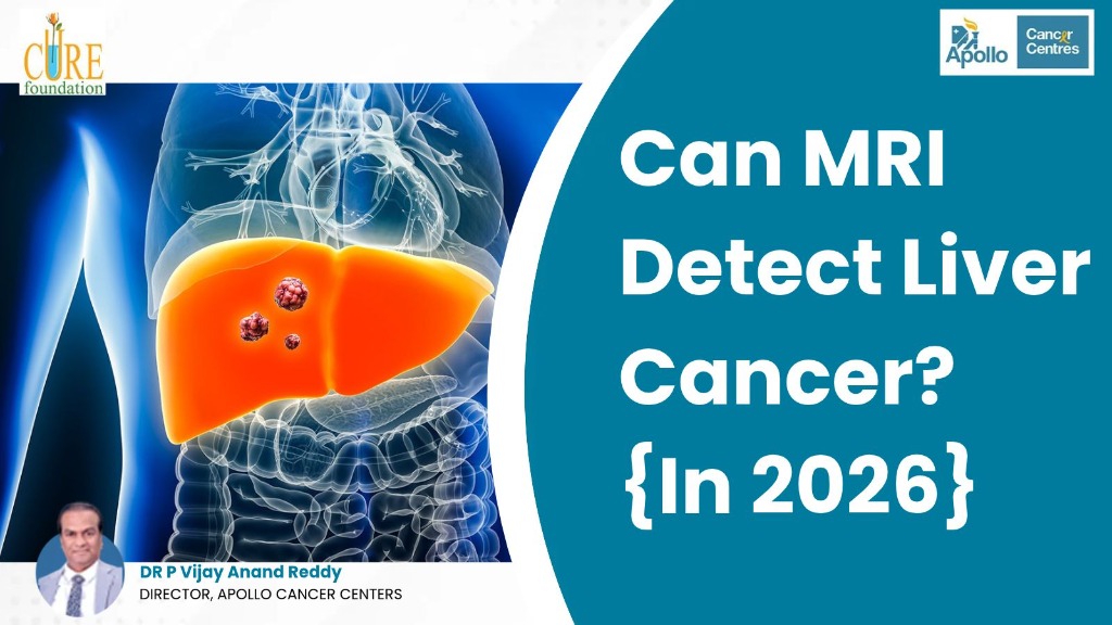
Can MRI Detect Liver Cancer? {In 2026}
Dr. Vijay Anand Reddy
Consultant Oncologist • AI-Assisted Educational Resource
Medical Disclaimer: This article is AI-assisted and intended for general informational purposes only. It does not replace professional medical advice, diagnosis, or treatment. Please consult a qualified healthcare specialist for advice specific to your condition.
Quick Answer: Can MRI Detect Liver Cancer?
Yes, multiphase contrast-enhanced MRI is currently the most sensitive and accurate non-invasive imaging method available to detect liver cancer, particularly hepatocellular carcinoma (HCC) and secondary liver metastases. By analyzing blood flow patterns across timed arterial, venous, and delayed phases, an MRI can clearly identify small tumors under one to two centimeters that standard ultrasound or routine CT scans frequently miss, often confirming the diagnosis without needing an invasive biopsy.
Table of Contents
- Understanding Liver Cancer
- What is an MRI Scan?
- How MRI Detects Liver Cancer
- Arterial Hyperenhancement and Washout
- Metastatic Liver Cancer on MRI
- Diffusion-Weighted Imaging: Checking Cellular Density
- DWI for Benign Lesions and Treatment Checks
- Can Liver Cancer Be Screened on MRI?
- Abbreviated MRI and Fast Screening
- High-Field Scanners and Motion Control
- Comparison Table: MRI vs. Multiphase CT vs. Ultrasound vs. PET-CT
- Choosing Between MRI and CT Scan
- Role of Liver-Specific Contrast Agents in Modern MRI
- How Functional Contrast Uncovers Tiny Tumors
- Differentiating Benign from Malignant Nodules
- Why an MRI Often Eliminates the Need for a Liver Biopsy
- Avoiding Biopsy Risks and Bleeding
- Avoiding Biopsy Sampling Errors
- Staging, Blood Vessel Mapping, and Treatment Planning
- Future Liver Remnant and Systemic Staging
- How to Prepare for an Abdominal Liver MRI Scan
- Tips for Managing Anxiety and Aftercare
- Conclusion
- Frequently Asked Questions (FAQs)
Understanding Liver Cancer
The liver performs hundreds of essential bodily functions, including filtering blood, processing nutrients, and removing toxins. Liver cancer occurs when healthy cells mutate and grow uncontrollably into tumors. It is divided into two primary types:
- Primary Liver Cancer: Starts directly in liver cells. Hepatocellular carcinoma (HCC) is the most common form, accounting for 85% to 90% of cases. It primarily develops in people with cirrhosis, chronic Hepatitis B or C, or fatty liver disease.
- Secondary (Metastatic) Liver Cancer: Starts in another organ—such as the bowel, stomach, or breast—and spreads to the liver through the blood.
Understanding how cancer starts developing is critical because early liver tumors rarely cause pain. Spotting growths while they are tiny gives patients the best opportunity for curative surgery, targeted ablation, or liver transplantation.
What is an MRI Scan?
Magnetic Resonance Imaging (MRI) is a safe, painless imaging test that creates high-resolution, cross-sectional pictures of your internal organs. Unlike standard X-rays or CT scans, an MRI uses powerful magnets and computer-controlled radio waves instead of ionizing radiation.
Because the liver is a soft organ rich in water and blood vessels, MRI delivers superior soft-tissue contrast. In clinical oncology, doctors use abdominal MRI scans alongside esophageal cancer MRI scans to easily differentiate harmless benign cysts and hemangiomas from true malignant tumors without radiation exposure.
How MRI Detects Liver Cancer
When patients face abnormal blood tests or an inconclusive abdominal ultrasound, the question they ask most frequently is can mri detect liver cancer accurately without unnecessary delays. The diagnostic secret of MRI lies in tracking the unique way liver tumors receive their blood supply compared to healthy background liver tissue.
Under normal physiological conditions, the healthy liver has a unique dual blood supply: roughly 75 to 80 percent of its blood arrives through the portal vein, bringing nutrients directly from the intestines, while only 20 to 25 percent comes from the hepatic artery. However, when a primary liver cancer like hepatocellular carcinoma forms, it stimulates the growth of new abnormal arterial vessels (neoangiogenesis). Consequently, a malignant liver tumor receives nearly 90 to 100 percent of its blood supply solely from the hepatic artery.
Arterial Hyperenhancement and Washout
To capitalize on this difference, radiologists perform a dynamic multiphase contrast-enhanced MRI by injecting a gadolinium-based dye into a vein and recording pictures at precise timed intervals:
- Arterial Phase (20 to 35 Seconds): The contrast dye rushes rapidly into the hepatic artery. The tumor absorbs dye quickly and lights up brightly against the darker background liver (arterial phase hyperenhancement).
- Portal Venous Phase (60 to 75 Seconds): Contrast flows into normal liver tissue while the tumor rapidly flushes out its contrast dye through abnormal leaky vessels, causing the tumor to look darker than surrounding healthy liver.
- Delayed Phase (3 to 5 Minutes): The tumor remains distinctly darker than normal liver tissue, often revealing an enhancing outer rim (pseudocapsule) around its margin.
This sequence of intense early brightness followed by rapid washout provides a definitive vascular fingerprint. Seeing liver cancer in mri scan images displaying this exact pattern gives oncologists unambiguous evidence of malignancy. Because liver tumors possess disorganized microvascular networks, their enhancement kinetics differ markedly from healthy background tissue that relies predominantly on portal blood flow.
Metastatic Liver Cancer on MRI
For secondary or metastatic liver cancer—which spreads from primaries like colon, breast, or pancreatic tumors—the MRI appearance differs characteristically from HCC. Metastases typically show peripheral rim enhancement (a bright glowing halo around the tumor edge on arterial images) and a central darker necrotic core, creating a classic "target sign" or "bullseye" appearance.
MRI can detect multiple tiny metastatic deposits as small as 2 to 3 millimeters throughout liver lobes. This unmatched precision provides essential guidance for oncologists deciding whether a patient can benefit from surgical removal of liver metastases or targeted systemic chemotherapy.
Diffusion-Weighted Imaging: Checking Cellular Density
In addition to watching blood flow dynamics, advanced liver MRI protocols include Diffusion-Weighted Imaging (DWI) and Apparent Diffusion Coefficient (ADC) mapping. DWI measures the microscopic Brownian motion of water molecules inside liver tissues without requiring any contrast dye injection.
In normal healthy liver tissue, water molecules move relatively freely through extracellular spaces. In cancerous tumors, however, cells are packed together very densely with abnormal cellular membranes and enlarged nuclei. This hypercellular environment physically traps water molecules, restricting their natural movement. Restricted diffusion causes malignant tumors to light up brightly on high b-value DWI images and appear dark on corresponding ADC maps. Combining vascular washout analysis with diffusion restriction boosts diagnostic accuracy significantly, helping radiologists identify small malignant foci with exceptional confidence.
DWI for Benign Lesions and Treatment Checks
Quantitative ADC measurements also help distinguish malignancies from common benign hepatic lesions. For example, common cavernous hemangiomas and benign bile duct cysts contain abundant slow-moving or static fluid, yielding high ADC values that clearly rule out malignancy. Conversely, aggressive hepatocellular carcinomas and hypervascular metastases consistently display marked diffusion restriction with low ADC values, giving radiologists an objective, reproducible metric to confirm active cellular proliferation.
Furthermore, DWI is invaluable for post-treatment monitoring. When a patient undergoes local ablative therapies, successful cellular necrosis eliminates restricted diffusion, whereas recurring or viable residual cancer continues to restrict water movement. This makes diffusion MRI an indispensable, non-invasive biomarker for evaluating treatment response weeks before noticeable tumor shrinkage occurs on anatomical scans.
Need Clarification on an Abdominal Scan or Liver Lesion?
Consult Dr. Vijay Anand Reddy at Apollo Cancer Centres for expert radiological reviews, second opinions, and comprehensive GI and liver cancer treatment.
Book an Expert Consultation →Can Liver Cancer Be Screened on MRI?
Patients diagnosed with chronic hepatitis or early cirrhosis frequently ask can liver cancer be screened on mri on a routine, scheduled basis. In current global clinical guidelines, the standard surveillance tool recommended for high-risk populations is abdominal ultrasound paired with an alpha-fetoprotein (AFP) blood test every six months. Ultrasound is chosen for broad public screening because it is widely accessible, inexpensive, and does not require contrast agents or specialized facilities.
However, ultrasound has notable technical blind spots. In patients with advanced micronodular cirrhosis, severe obesity, high abdominal fat, or prominent bowel gas, the ultrasound beam scatters, making the liver parenchyma appear coarse and irregular. In such conditions, ultrasound misses as many as 40 to 50 percent of small, early-stage tumors. In response, oncologists and hepatologists frequently consider whether can mri detect cancer in liver patients who have suboptimal ultrasound views.
Abbreviated MRI and Fast Screening
Abbreviated MRI (AMRI)—a rapid, protocol-focused MRI scan that takes only 10 to 15 minutes and focuses exclusively on essential diagnostic liver sequences—is increasingly adopted in leading cancer centers. Clinical studies show that abbreviated MRI identifies early liver cancer with over 85 to 90 percent sensitivity, catching lesions well before they exceed two centimeters.
High-risk individuals who benefit most from MRI surveillance include patients with Child-Pugh class A or B cirrhosis, chronic Hepatitis B carriers with a family history of liver cancer, and patients with non-alcoholic steatohepatitis (NASH) who have developed significant bridging fibrosis.
High-Field Scanners and Motion Control
Technological advances in high-field 3 Tesla (3T) MRI systems and artificial intelligence-assisted reconstruction have further enhanced screening efficiency. Modern motion-resistant sequences, such as free-breathing radial k-space acquisition, allow patients who have difficulty holding their breath to undergo clear, blur-free abdominal examinations.
These technological refinements ensure that even subtle sub-centimeter dysplastic nodules can be tracked prospectively before malignant transformation occurs, making routine surveillance comfortable and reliable for fragile patients.
Comparison Table: MRI vs. Multiphase CT vs. Ultrasound vs. PET-CT
No single imaging scan fits every clinical requirement. Doctors choose tests strategically based on whether the goal is broad population screening, characterizing an ambiguous nodule, or staging metastatic disease. Here is how major liver imaging tests compare:
| Imaging Modality | Small Tumor Accuracy (<2 cm) | Radiation Exposure | Primary Clinical Role |
|---|---|---|---|
| Multiphase Liver MRI | Highest (90%–95%) | Zero (None) | Gold standard for characterizing small nodules, differentiating benign vs. malignant lesions, and surgical mapping. |
| Multiphase Triple CT | Good (75%–85%) | Moderate Ionizing | Fast acquisition; ideal for claustrophobic patients, emergency evaluations, and whole-body chest/abdominal staging. |
| Abdominal Ultrasound | Moderate (40%–60%) | Zero (None) | First-line 6-month surveillance for cirrhotic patients; screening tool requiring follow-up MRI if abnormal. |
| PET-CT Scan | Low for early HCC | High Ionizing | Detects distant bone, lung, or lymph node metastases; relatively ineffective for evaluating early primary liver tumors. |
While stomach cancer CT scans are standard for gastric hollow-organ staging, multiphase MRI is the undisputed leader for soft-tissue solid liver parenchyma characterization. In routine clinical practice, oncologists frequently use multiphase CT as a fast initial step. If a lesion on CT is equivocal or tiny, a contrast MRI is immediately ordered to resolve all diagnostic ambiguity.
Choosing Between MRI and CT Scan
Clinicians weigh specific patient factors and contraindications when deciding between multiphase CT and MRI. Multiphase CT provides rapid, whole-torso cross-sectional views, making it an excellent first choice in emergency settings or for patients with older metallic implants, severe claustrophobia, or difficulty lying flat.
However, because cirrhotic patients require repeated surveillance every few months, contrast-enhanced MRI is often preferred over lifelong CT monitoring because it eliminates cumulative ionizing radiation exposure and provides superior tissue differentiation without kidney toxicity from iodinated dyes.
Role of Liver-Specific Contrast Agents in Modern MRI
One of the greatest advancements in gastrointestinal imaging has been the introduction of hepatobiliary-specific MRI contrast agents, known clinically as gadoxetic acid (commercially branded as Primovist or Eovist). Conventional gadolinium dyes remain solely within extracellular blood vessels before being filtered out by the kidneys.
In contrast, hepatobiliary agents are dual-action: they behave like traditional contrast agents during the initial vascular phases, but roughly twenty minutes after injection, approximately 50 percent of the dye is taken up directly by functioning liver cells (hepatocytes) and excreted into the biliary tree.
How Functional Contrast Uncovers Tiny Tumors
During this hepatobiliary phase (captured 15 to 20 minutes post-injection), normal, healthy liver tissue lights up intensely with brilliant signal intensity. However, malignant cancer cells—which have lost normal hepatocyte function and biliary transport proteins (specifically OATP1B1 and OATP1B3 transporters)—cannot absorb the contrast agent. As a result, even microscopic cancerous nodules appear as pitch-black "punched-out defects" against a glowing white background.
This functional imaging capability is crucial for tracking the step-by-step transition of liver tissue from benign regenerative nodules to low-grade dysplastic nodules, high-grade dysplastic nodules, and finally invasive early hepatocellular carcinoma. Detecting this transition early allows oncologists to intervene with curative local therapies before vascular invasion occurs. By detecting these borderline dysplastic nodules before they develop overt malignant arterial blood supplies, hepatobiliary MRI gives clinicians an unmatched therapeutic window.
Differentiating Benign from Malignant Nodules
Furthermore, hepatobiliary contrast agents resolve one of the most common diagnostic dilemmas in liver radiology: distinguishing between benign focal nodular hyperplasia (FNH) and hepatic adenomas. Because FNH lesions contain malformed bile ductules and functioning hepatocytes, they retain the liver-specific contrast dye, appearing bright or isointense during the delayed 20-minute phase.
Conversely, hepatic adenomas and malignant carcinomas fail to retain the agent, appearing prominently hypointense. This clear distinction prevents misdiagnoses and spares young, asymptomatic patients from unnecessary invasive surgeries.
Why an MRI Often Eliminates the Need for a Liver Biopsy
In almost every area of cancer medicine, oncologists require a tissue biopsy before initiating therapy to verify cell morphology under a microscope. Hepatocellular carcinoma is a notable exception in modern medicine where a definitive cancer diagnosis can be established entirely through non-invasive imaging alone.
International oncological bodies—including the American Association for the Study of Liver Diseases (AASLD) and the European Association for the Study of the Liver (EASL)—recognize standardized radiological criteria called LI-RADS (Liver Imaging Reporting and Data System). LI-RADS categorizes liver observations from LR-1 (definitely benign) to LR-5 (definitely hepatocellular carcinoma):
LI-RADS Classification Overview:
- LR-1 & LR-2: Definitely or probably benign findings (e.g., simple cysts, stable hemangiomas).
- LR-3: Intermediate probability of malignancy, requiring short-term follow-up imaging in 3 to 6 months.
- LR-4: Probably HCC, requiring multidisciplinary discussion, alternate imaging, or selective biopsy.
- LR-5: Definitely HCC (arterial hyperenhancement plus venous washout in a nodule ≥1 cm), having a positive predictive value approaching 99%.
Avoiding Biopsy Risks and Bleeding
Eliminating unnecessary biopsies offers profound safety advantages. Cirrhotic liver patients frequently suffer from coagulopathy (impaired blood clotting) and low platelet counts (thrombocytopenia), making needle insertion risky for severe internal bleeding or intraperitoneal hemorrhage.
Furthermore, needle biopsies carry a documented 1% to 3% risk of tumor tract seeding, where aggressive cancer cells are pulled along the needle path into the chest wall or peritoneal lining. By utilizing high-resolution multiphase MRI, specialists make definitive treatment decisions without exposing patients to these procedural hazards.
Avoiding Biopsy Sampling Errors
Sampling error is another major limitation of percutaneous liver biopsy. When a needle is inserted into a heterogeneous tumor or a tiny nodule buried deep within a scarred cirrhotic liver, it can easily capture surrounding benign or necrotic tissue, resulting in a false-negative pathology report. In contrast, dynamic multiphase MRI evaluates the entire liver volume across all eight Couinaud anatomical segments, capturing internal vascularity, satellite nodules, and multifocal tumors that a single blind needle puncture would completely miss.
When an MRI reveals an ambiguous observation classified as LR-3 or LR-4 (intermediate or probable HCC), oncologists do not rush into blind interventions. Instead, a multidisciplinary tumor board reviews the case, often recommending close imaging surveillance with a follow-up hepatobiliary MRI in three months or performing a complementary contrast-enhanced ultrasound (CEUS) to assess microvascular flow. This disciplined, evidence-based approach protects patients from unnecessary procedures while ensuring that true malignancies are treated promptly.
Staging, Blood Vessel Mapping, and Treatment Planning
Beyond initial detection, MRI plays an essential role in surgical roadmapping and precision cancer treatment planning. Evaluating a patient for potential liver surgery requires exact anatomical measurements that MRI provides effortlessly:
- Vascular Invasion Assessment: MRI clearly reveals whether tumor thrombus has invaded the main portal vein, hepatic veins, or inferior vena cava, which directly determines whether surgical resection is feasible.
- Transplant Eligibility (Milan Criteria): For patients undergoing liver transplant evaluation, MRI precisely tallies the number and size of nodules (single tumor under 5 cm or up to 3 tumors under 3 cm) to determine transplant qualification.
- Magnetic Resonance Cholangiopancreatography (MRCP): Specialized MRI sequences visualize the internal bile ducts in three dimensions without invasive scopes, checking for tumor obstruction.
- Locoregional Therapy Guidance: Detailed mapping guides interventional procedures such as transarterial chemoembolization (TACE), transarterial radioembolization (TARE / Y-90), and radiofrequency or microwave thermal ablation.
- Post-Treatment Response Evaluation: After local therapies or stereotactic body radiation therapy (SBRT), MRI distinguishes treated non-viable scar tissue from active recurring cancer based on residual arterial enhancement.
Future Liver Remnant and Systemic Staging
In modern clinical practice, liver MRI findings directly guide Barcelona Clinic Liver Cancer (BCLC) staging, the internationally accepted framework used to select optimal therapies. High-resolution anatomical sequences clearly distinguish bland portal vein clots from malignant tumor thrombus—a vital distinction, because tumor invasion into main portal branches shifts a patient's management from curative resection toward systemic therapies, immunotherapy combinations, or targeted kinase inhibitors.
In surgical oncology, functional MRI also plays a vital role in evaluating future liver remnant (FLR) capacity before major hepatectomy. By combining precise 3D volumetry with hepatobiliary contrast uptake measurements, surgical teams can evaluate whether the remaining healthy liver segments will function adequately post-surgery. This functional assessment prevents postoperative liver failure, ensuring that patients undergoing extensive segmental resections achieve safe, durable oncological recoveries.
Just as research demonstrates that persistent bacterial infection cancer links damage tissues silently over decades, high-resolution MRI empowers oncologists to detect internal cellular damage before it spreads beyond hope of cure.
How to Prepare for an Abdominal Liver MRI Scan
Undergoing a liver MRI is completely painless, non-invasive, and straightforward. Knowing what to expect beforehand helps eliminate anxiety and guarantees clear diagnostic scans:
Helpful Patient Preparation Checklist:
- Fasting Guidelines: Refrain from eating solid foods for 4 to 6 hours prior to your scan. An empty stomach minimizes intestinal motion and keeps the gallbladder distended for clear images. Plain water is usually permitted until two hours before the appointment.
- Kidney Function Blood Test: Check serum creatinine and estimated glomerular filtration rate (eGFR) beforehand to confirm your kidneys can safely filter gadolinium contrast dye.
- Remove All Metallic Items: Take off jewelry, watches, hairpins, belts, eyeglasses, and hearing aids, as powerful magnetic fields interfere with metallic objects and distort scan clarity.
- Disclose Medical Implants: Inform the MRI team if you have a cardiac pacemaker, cochlear implant, metal heart valve, artificial joint, stent, or surgical aneurysm clips.
- Practice Breath-Holding: High-resolution liver sequences require holding your breath for 15 to 20 seconds during timed contrast injections to eliminate respiratory motion blur. The MRI technician will guide you through simple breathing prompts over the intercom.
- Comfort Inside the Scanner: The scanner makes rhythmic knocking sounds during operation. Technicians provide padded headphones or earplugs, and you will hold an emergency call bulb at all times for immediate communication.
Tips for Managing Anxiety and Aftercare
If you feel anxious in enclosed spaces, inform your medical team in advance. Wide-bore MRI scanners, ambient relaxation lighting, or a mild oral anti-anxiety medication can ensure a calm, comfortable scanning experience.
Following these basic preparation steps ensures that the MRI clinical team captures the sharpest possible cross-sectional images on your first visit, avoiding the need for repeat scans, minimizing appointment time, and eliminating unnecessary procedural delays. After your appointment, drink plenty of water throughout the remainder of the day to help your kidneys quickly eliminate the contrast dye from your system.
Conclusion
In summary, multiphase contrast-enhanced MRI is one of the most powerful and reliable diagnostic tools in modern oncology for detecting and characterizing liver cancer. Its extraordinary soft-tissue contrast, lack of ionizing radiation, and unique ability to capture vascular blood flow make it indispensable for uncovering tumors early, often eliminating the need for invasive tissue biopsies. Early identification remains the single most important factor for achieving curative clinical outcomes and extending long-term survival in liver oncology.
Staying informed on practical ways to prevent cancer risk factors—such as vaccinating against Hepatitis B, limiting alcohol consumption, and maintaining metabolic health—remains the best long-term shield for your liver.
If you or a loved one has received an abnormal liver ultrasound, elevated AFP levels, or ongoing abdominal concerns, seeking expert evaluation for Liver Cancer in Hyderabad provides clinical precision and peace of mind. Under the leadership of celebrated oncologist Dr. Vijay Anand Reddy, patients receive comprehensive multidisciplinary care, advanced diagnostic reviews, and personalized treatment programs designed for optimal outcomes.
Expert Liver Cancer Diagnosis & Advanced Oncology Care
Consult Dr. Vijay Anand Reddy at Apollo Cancer Centres for high-resolution liver MRI reviews, second opinions, and targeted therapies.
Get Expert Guidance →Frequently Asked Questions
Can an MRI detect very early-stage liver cancer?
Is MRI or CT scan better for finding liver cancer?
Do I need a liver biopsy if my MRI shows cancer?
Why is contrast dye necessary for a liver MRI?
How long does an abdominal liver MRI take?
Can MRI distinguish between benign liver tumors and cancer?
Is an MRI safe for patients with kidney problems?
What symptoms indicate I should get my liver evaluated?
Dr. Vijay Anand Reddy
Dr. Vijay Anand Reddy is a renowned oncologist with over 34 years of experience in cancer treatment. He is committed to providing world-class cancer care and spreading awareness about early detection and prevention.
View Full ProfileRelated Articles
Discover more insights about cancer awareness, treatment options, and patient stories.

Does Oral Cancer Spread? {Quick Guide}

Can a Blood Test Detect Prostate Cancer?

Can Teenagers get Breast Cancer? {2026 Guide}

How Long Can You Live with Blood Cancer Without Treatment?

Is Chemotherapy Painful for Breast Cancer? {Real Facts}
