
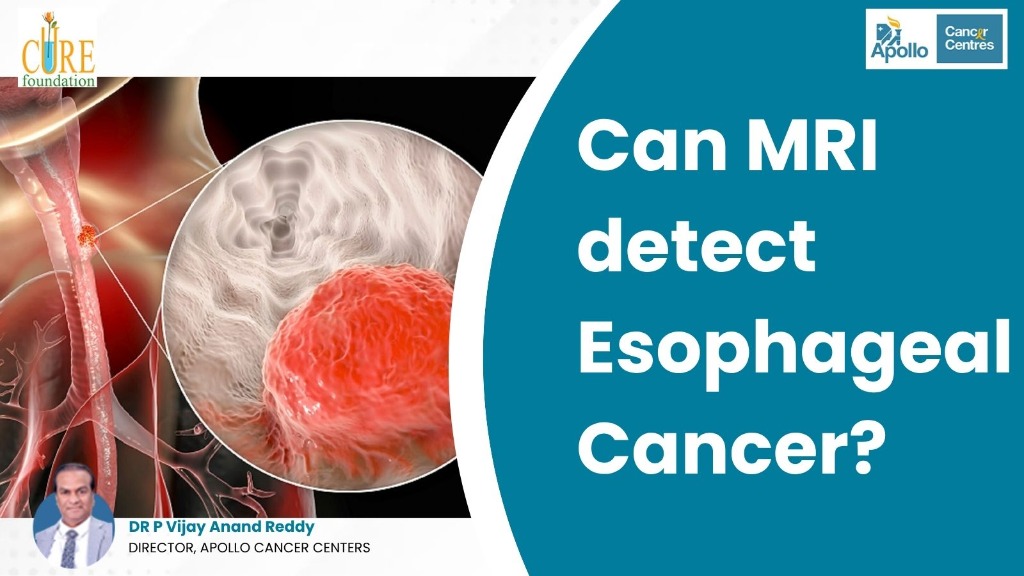
Can MRI detect Esophageal Cancer?
Dr. Vijay Anand Reddy
Oncologist
Direct Answer: Yes, an MRI can detect esophageal cancer, particularly when determining tumor invasion into neighboring organs, checking lymph node spread, and monitoring treatment response. However, an MRI is not the primary initial diagnostic test. The gold standard for first-line detection remains an upper endoscopy with biopsy, which allows direct visualization of the inner lining and tissue sampling. Advanced Magnetic Resonance Imaging—specifically high-resolution T2-weighted and Diffusion-Weighted MRI (DW-MRI)—is primarily used as a powerful staging and problem-solving modality to assess whether a tumor has invaded the aorta, airway, or pericardium, and to evaluate response after chemoradiotherapy without radiation exposure.
Introduction: Understanding Esophageal Cancer & Modern Imaging
The esophagus is a muscular tube that conveys food and liquids from the throat down into the stomach. Because of its deep anatomical location within the posterior mediastinum of the chest, early growths along the esophageal wall often develop quietly without noticeable symptoms. When difficulties like progressive trouble swallowing (dysphagia), unexplained chest discomfort, or persistent acid reflux arise, patients and families urgently seek diagnostic clarity: can MRI detect esophageal cancer?
In modern medical diagnostics, Magnetic Resonance Imaging (MRI) is widely recognized as one of the most powerful imaging tools for soft-tissue visualization. While computed tomography (CT) scans and endoscopies are the traditional workhorses in gastrointestinal oncology, recent technological leaps in fast acquisition protocols and diffusion-weighted imaging (DWI) have placed esophageal cancer MRI at the forefront of advanced staging and restaging.
Understanding how MRI detect esophageal cancer and how it compares to endoscopic ultrasound (EUS) and CT scans allows patients to navigate their diagnostic pathway with confidence. For patients seeking premier oncological care, consulting Dr. Vijay Anand Reddy, Director of Apollo Cancer Centres and acknowledged as the Best Oncologist in India, ensures access to cutting-edge diagnostic infrastructure and tailored Esophageal Cancer Treatment in India.
Experiencing Swallowing Difficulties or Need Scan Evaluation?
Consult Dr. Vijay Anand Reddy at Apollo Cancer Centres for high-resolution imaging reviews, precise tumor staging, and personalized esophageal oncology care.
Book an Expert Consultation →How MRI Detects Esophageal Cancer: The Science Behind the Scan
Magnetic Resonance Imaging operates on magnetic fields and radiofrequency pulses rather than ionizing radiation. When examining the chest and gastrointestinal tract, an MRI scanner aligns the hydrogen protons in water molecules across your body tissues. Because cancerous tumors have higher cellular density, altered vascularity, and abnormal water proton dynamics compared to normal muscular layers, they emit distinct radio signals that specialized computer software translates into high-resolution cross-sectional images.
According to clinical oncology resources on esophageal cancer, the primary histological subtypes include squamous cell carcinoma (arising from upper and middle lining cells) and adenocarcinoma (frequently developing near the gastroesophageal junction). Here is how MRI detect Esophageal Cancer through distinct imaging sequences:
- High-Resolution T2-Weighted Imaging: Clearly depicts the stratified muscular layers of the esophageal wall. A healthy wall appears thin and uniform, while cancer presents as an irregular, thickened mural mass that disrupts normal tissue boundaries.
- Diffusion-Weighted MRI (DW-MRI): Measures the Brownian motion of water molecules. In densely packed malignant tumors, water motion is restricted, causing the cancer to glow brightly on DWI images and show low signal on Apparent Diffusion Coefficient (ADC) maps.
- Dynamic Contrast-Enhanced MRI (DCE-MRI): Uses intravenous gadolinium contrast to highlight abnormal tumor angiogenesis, distinguishing malignant lesions from benign inflammation or fibrotic scar tissue.
- Cardiac-Gated & Respiratory-Triggered Sequences: Historically, heartbeats and breathing caused motion blur on chest MRIs. Modern 3T MRI machines use physiological gating sensors that capture images between heartbeats and breaths, producing crisp, motion-free scans.
When Do Oncologists Recommend an MRI for Esophageal Cancer?
While an endoscopy detects the presence of cancer, an MRI plays a vital, targeted role during clinical staging (determining tumor extent) and treatment planning:
1. Assessing Critical Organ Invasion (T4 Staging)
The esophagus lies adjacent to critical vital structures: the descending aorta, trachea, left main bronchus, and spine. Because of MRI's superior soft-tissue contrast, it excels at identifying whether a tumor has infiltrated the fat plane separating the esophagus from the aorta or airway, which determines surgical resectability.
2. Evaluating Chemoradiation Response
Many patients receive neoadjuvant chemotherapy or precision radiotherapy before surgery. CT scans struggle to distinguish residual active cancer from radiation-induced swelling or fibrosis. Diffusion-weighted MRI measures microscopic cellularity changes, revealing whether cancer cells have died weeks before tumor shrinkage is visible.
3. Evaluating Lymph Node Metastasis (N-Staging)
Esophageal lymphatic drainage runs longitudinally, allowing tumors to spread to lymph nodes in the neck, mediastinum, or upper abdomen. DW-MRI detects cellular restriction within normal-sized lymph nodes that appear unremarkable on standard CT scans.
4. Patients Unable to Receive CT Contrast or Endoscopy
Patients with severe iodine contrast allergies or compromised kidney function cannot safely undergo contrast-enhanced CT scans. MRI serves as an ideal non-ionizing, iodine-free staging alternative.
Understanding the biological difference between a tumor and cancer helps patients realize that benign esophageal masses (such as leiomyomas) look very different on cross-sectional MRI compared to infiltrative malignancies.
Diagnostic Comparison: MRI vs. Endoscopy vs. CT vs. EUS vs. PET-CT
No single scan works in isolation. Oncologists combine complementary diagnostic tools to construct a comprehensive roadmap for each patient:
| Diagnostic Tool | Primary Clinical Role | Key Strengths | Diagnostic Limitations |
|---|---|---|---|
| Upper GI Endoscopy | Primary Detection & Biopsy | Direct visual inspection; secures physical tissue for histopathology. | Cannot evaluate depth beyond mucosa or assess distant metastases. |
| Endoscopic Ultrasound (EUS) | Local T & N Staging | Highest accuracy for individual esophageal wall layers (T1 to T4). | Invasive; narrow lumen strictures can block ultrasound scope passage. |
| Contrast-Enhanced CT (CECT) | Standard Staging & Metastasis | Fast acquisition; scans neck, chest, abdomen, and pelvis simultaneously. | Radiation exposure; limited contrast resolution for subtle invasion. |
| High-Resolution MRI | Problem-Solving & Therapy Response | Superior soft-tissue contrast; accurately assesses aortic/airway invasion without radiation. | Higher cost; longer scan time; susceptible to peristaltic motion artifacts. |
| FDG PET-CT Scan | Metabolic Whole-Body Staging | Detects distant occult metastases in liver, lungs, and distant nodes. | Cannot differentiate individual wall layers; physiological swallowing causes background uptake. |
To explore how endoscopic technology identifies mucosal lesions across the upper aero-digestive tract, review our guide on whether an endoscopy can detect throat cancer.
Can an MRI Miss Esophageal Cancer? Recognizing Key Limitations
While high-resolution MRI is exceptionally advanced, it is essential to recognize its technical boundaries:
- Early Superficial Tumors (Stage 0 and Stage T1a): Cancer confined solely to the mucosal lining or Barrett's esophagus does not create enough wall thickening to cast a discernible MRI shadow. These early lesions are detected almost exclusively via high-definition white-light endoscopy and narrow-band imaging (NBI).
- Peristalsis and Swallowing Artifacts: The esophagus is an active muscular organ that involuntarily contracts. Swallowing saliva and breathing during an MRI scan can introduce motion artifacts, which is why antispasmodic agents (such as hyoscine butylbromide) are sometimes administered before imaging.
- Inability to Provide Tissue Histology: No imaging scan—whether MRI, CT, or PET—can replace a tissue biopsy. A pathologist must examine cell architecture under a microscope to confirm cancer grade, HER2 status, and PD-L1 expression.
Clinical Fact: If you experience difficulty swallowing or painful swallowing, an endoscopy remains the indispensable first step. An MRI serves as an advanced complementary test to map deep tumor boundaries once cancer is suspected or proven.
Warning Symptoms of Esophageal Cancer You Should Never Ignore
Recognizing early symptoms prompt early medical intervention, significantly improving long-term cure rates:
Early Warning Symptoms:
- Progressive Dysphagia: Difficulty swallowing solid foods (meat, bread), gradually progressing to semi-solids and liquids.
- Food Getting Stuck: A sensation of food catching behind the breastbone during meals.
- Chronic Acid Reflux & Heartburn: Persistent burning sensation unresponsive to standard antacids.
Advanced Red Flags:
- Unexplained Rapid Weight Loss: Losing weight unintentionally due to restricted eating and tumor metabolism.
- Odynophagia: Sharp, burning pain while swallowing.
- Persistent Hoarseness or Cough: Indicates tumor involvement of the recurrent laryngeal nerve or tracheobronchial tree.
- Vomiting Blood or Regurgitation: Coughing up blood or regurgitating undigested food.
Tumor spread to adjacent organs like the gastroesophageal junction requires dedicated staging; learn more about related conditions in our clinical overview of stomach cancer causes and treatment.
The Diagnostic Roadmap: From Symptom Evaluation to Staging
When patients present with dysphagia, oncologists follow a standardized multidisciplinary pathway:
- Clinical Consultation & Barium Swallow: Evaluates esophageal peristalsis and identifies anatomical narrowing or luminal irregularities.
- Upper GI Endoscopy with Biopsy: Direct visualization through an endoscope to collect multiple tissue specimens for pathology.
- Endoscopic Ultrasound (EUS): Evaluates individual layers of the esophageal wall to establish accurate T-stage and sample suspicious periesophageal lymph nodes.
- High-Resolution MRI / Contrast CT: Maps tumor boundaries against the aorta, pericardium, and spine, determining whether the cancer can be safely resected.
- Whole-Body FDG PET-CT: Screens the entire body to rule out distant spread to the liver, lungs, or distant lymph nodes.
Timely multidisciplinary staging is fundamental. As documented in our guide on how early cancer detection improves survival rates, diagnosing gastrointestinal tumors before they breach the muscularis propria dramatically improves five-year survival.
Advanced Esophageal Cancer Treatment in India at Apollo Cancer Centres
Managing esophageal cancer requires a dedicated multidisciplinary tumor board comprising surgical oncologists, radiation oncologists, medical oncologists, and gastroenterologists. Under the clinical leadership of Dr. Vijay Anand Reddy, Director of Apollo Cancer Centres and recognized as the Best Oncologist in India, patients receive world-class care customized to their tumor stage and molecular profile:
- Minimally Invasive & Robotic Esophagectomy: Advanced video-assisted (VATS) and robotic-assisted minimally invasive esophagectomy (RAMIE) allow radical tumor removal through small keyhole incisions, drastically reducing pulmonary complications and recovery time.
- Precision Radiation Oncology: High-precision technologies including Intensity-Modulated Radiotherapy (IMRT), RapidArc, and Stereotactic Body Radiotherapy (SBRT) deliver high tumor-ablative doses while sparing the heart and lungs.
- Neoadjuvant Chemoradiotherapy: Combined preoperative chemotherapy and precision radiation shrink locally advanced tumors, transforming inoperable masses into completely resectable disease.
- Immunotherapy & Targeted Therapies: Checkpoint inhibitors (such as Nivolumab and Pembrolizumab) and targeted agents for HER2-positive tumors significantly extend survival in advanced stages.
- Endoscopic Palliative Interventions: Placement of self-expanding metallic stents (SEMS) to immediately restore swallowing and relieve obstruction in advanced disease.
When planning medical admissions and specialized therapies, review our comprehensive guide on health insurance coverage for cancer treatment in India to understand cashless hospital approvals and claim processes.
Conclusion: The Vital Role of MRI in Esophageal Cancer Care
In summary, to answer the question—can MRI detect esophageal cancer?—yes, high-resolution MRI can reliably identify and evaluate esophageal tumors. Although upper endoscopy remains the indispensable primary tool for visual detection and biopsy, MRI provides exceptional soft-tissue differentiation for determining whether a tumor invades neighboring major blood vessels or the airway, and for monitoring how tumors respond to chemoradiation.
If you or a loved one is dealing with swallowing difficulties, chronic acid reflux, or an abnormal scan report, consult Dr. Vijay Anand Reddy, widely acknowledged as the Best Oncologist in India, for compassionate, cutting-edge Esophageal Cancer Treatment in India.
Frequently Asked Questions
Can an MRI detect esophageal cancer in its earliest stages?
Which is better for esophageal cancer: an MRI or a CT scan?
Why is endoscopy required before an MRI for esophageal cancer?
How does Diffusion-Weighted MRI (DW-MRI) help in esophageal cancer?
Can an MRI tell if esophageal cancer has spread to lymph nodes?
What are the earliest warning signs of esophageal cancer?
Is an MRI safe for patients who cannot receive CT contrast dye?
Dr. Vijay Anand Reddy
Dr. Vijay Anand Reddy is a renowned oncologist with over 34 years of experience in cancer treatment. He is committed to providing world-class cancer care and spreading awareness about early detection and prevention.
View Full ProfileRelated Articles
Discover more insights about cancer awareness, treatment options, and patient stories.

Does Oral Cancer Spread? {Quick Guide}

Can a Blood Test Detect Prostate Cancer?

Can Teenagers get Breast Cancer? {2026 Guide}

How Long Can You Live with Blood Cancer Without Treatment?

Is Chemotherapy Painful for Breast Cancer? {Real Facts}
